Lamb mortality in the first month of life
With the bulk of flocks getting through their lambing, and the weather not really improving, losses in the first month of life may well be higher this year than most. Gross postmortem examination can provide a quick answer for farmers, with diagnostic testing used to support the diagnosis. Below is a quick review of the postmortem findings and diagnostic tests available for some of the most common causes of sudden death after turnout.
- Pneumonia/Pasteurellosis
- Colisepticaemia
- Pulpy kidney
- Lamb dysentery
- Intestinal torsion
- Starvation
Pneumonia/Pasteurellosis
Death can be preceded by respiratory signs, but lambs are most often found dead. In lambs under 1 month old Mannheimia haemolytica is by far the most common cause, however Bibersteinia trehalosi or Pasteurella multocida can also affect this age group.
The gross findings can be variable:
- Cranioventral consolidation (classic presentation)
- Fibrinous pleurisy (often associated with M. haemolytica)
- Multifocal abscesses within the lung parenchyma (commonly due to Trueperella pyogenes)
- Diffuse, patchy, dark discolouration of lung lobules (acute infection/septicaemia, often M. haemolytica)
Diagnosis of the causal organisms can be done by bacterial culture, PCR testing (not validated) and histopathology of the affected tissue.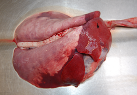
Colisepticaemia (watery mouth)
Escherichia coli septicaemia is most commonly diagnosed in lambs under one week old, and is associated with inadequate colostrum intake. Diagnosis is surprisingly challenging as there are often no gross postmortem findings. There is also often little to be seen on histopathology, although signs of toxaemia can be seen in some cases. Culture of profuse, pure growth of E. coli from multiple systemic sites (e.g. liver, spleen, kidney) is supportive, although care must be taken to avoid contamination with intestinal contents or faeces. As E. coli is a commensal of the GI tract, abomasal culture is not diagnostically useful. Collection of blood at postmortem exam (or from cohort animals) and demonstration of a low ZST test result indicating failure of passive transfer is also supportive.
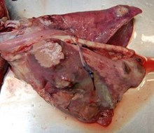
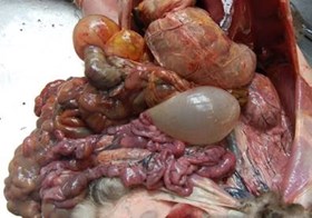
Pulpy kidney
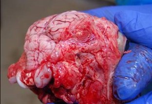
Most diagnoses of pulpy kidney (Clostridium perfringens type D disease) occur in April and May. A small number of these diagnoses are in neonatal animals, however this may be underdiagnosed since postmortem examination of this age of lamb is less common.
The presentation of neonatal disease often features neurological signs such as ataxia, recumbency, seizures and muscle spasticity reported prior to death, as well as sudden death. The gross findings of postmortem examination are similar to older animals with an excess of pericardial fluid +/- fibrin clot (pictured below), pulmonary oedema, and cerebellar coning (pictured below) common in affected lambs. Advanced autolysis in the kidneys compared to the rest of the carcase is a less common finding in neonatal lambs. Diagnosis is by detection of epsilon toxin in the terminal ileal content and/or focal symmetrical encephalomalacia on histopathological examination of the fixed brain.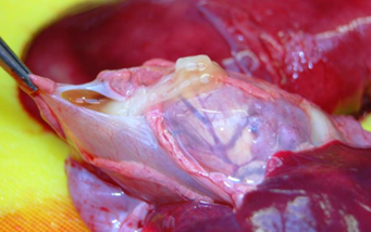
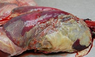
Lamb dysentery
We are now entering the period where lamb dysentery (Clostridium perfringens type B disease) risk is highest in the main April lambing flock. Most commonly seen in outdoor lambing flocks and presenting as sudden death, occasionally with reports of a preceding bloody scour. Traditionally thought of as a disease in very young lambs the majority of diagnoses are made in lambs over seven days of age.
In fresh carcasses there is often a characteristic emphysema of the jejunal serosa (pictured below), however this can be lost with autolysis. There is often petechiation of the serosal surface of affected intestine, which can also be darkly congested. Demonstration of beta and/or epsilon toxins from terminal ileal content is the diagnostic method of choice and can be supported by histopathology of intestine.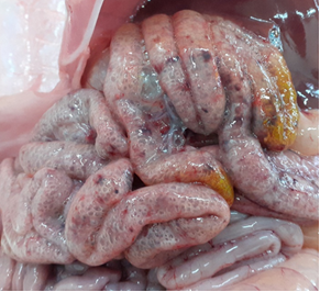
Intestinal torsion
Also known as red gut, intestinal torsions are sporadic, and more often seen in well grown or hand-reared lambs, especially associated with the introduction of creep feed. It is thought that fermentation of carbohydrate within the intestine leads to gassy distention and displacement, with intestines usually rotating around the root of the mesentery. On postmortem exam there is usually gassy distension of the abdomen/intestine, commonly with dark serosal discolouration with prominent vascular congestion in the mesentery and intestinal serosa, and dark bloody contents within the affected gut. Often the torsion has corrected itself at the time of PM exam. The diagnosis is made on gross postmortem exam.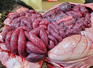
Starvation
Commonly due to mismothering or rejection, which is most common in lambs under one month old. On external examination lambs are typically small, poorly muscled with sunken eyes (dehydration), meaning depletion of body fat and tacky skin/viscera are common observed on gross postmortem. An empty abomasum and scant contents in GI tract in younger lambs is supportive of the diagnosis. In two to four week old lambs, the rumen is commonly full of fibrous content with no or very little milk in the abomasum - the underdeveloped rumen means that they cannot survive on grass only at this age. Some cases occur secondary to joint-ill therefore examination of the joints (with bacterial culture of any abnormal joints) is worthwhile. Diagnosis is made on gross examination.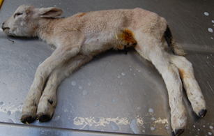
Posted by SRUC Veterinary Services on 23/04/2024